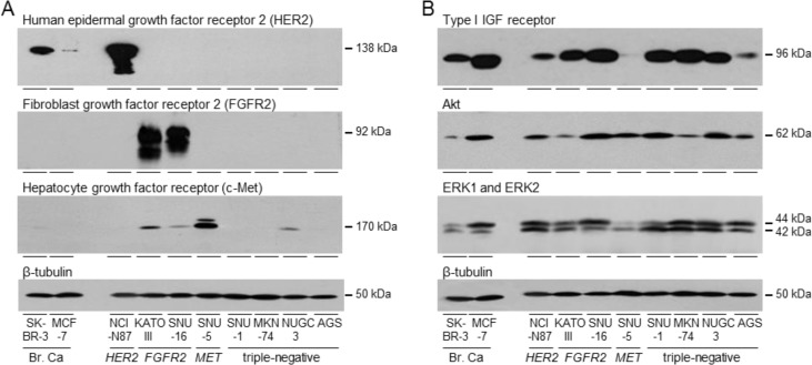
Figure 1. Expression of tyrosine kinase receptors in gastric cancer cells.
SK-BR-3, MCF-7, NCI-N87, KATO III, SNU-16, SNU-5, SNU-1, MKN74, NUGC3 and AGS were grown to 70% confluence and lysed. Aliquots of 10 μg of protein were electrophoresed on polyacrylamide gels, transferred to nitrocellulose and incubated with antibodies against HER2, FGFR2, c-Met A., type I IGF receptor, pan Akt, ERK1 and ERK2 B. and β-tubulin and developed as described in the Materials and Methods. Representative images are shown.

