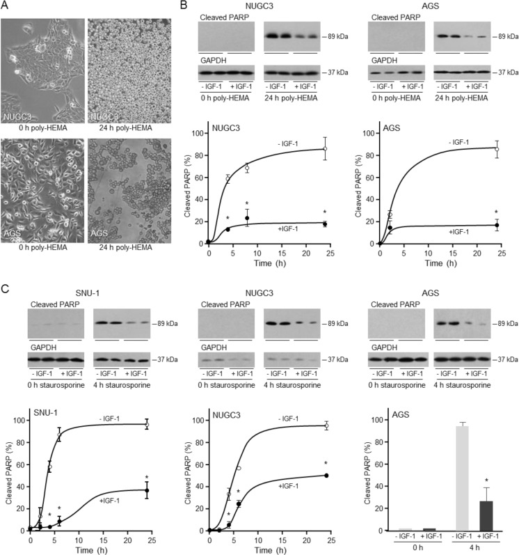
Figure 3. Protective effect of IGF-1 against anoikis and apoptosis in triple-negative gastric cancer cells.
For induction of anoikis, NUGC3 and AGS cells were added to uncoated or poly-HEMA-coated plates A. or to poly-HEMA-coated plates in the absence or presence of 50 ngml−1 IGF-1 B. Cells cultured in poly-HEMA-coated plates lost their characteristic polygonal appearance and grew as rounded detached cells A. Cells were lysed and the amount of cleaved PARP was analysed by Western transfer B. The amount of cleaved PARP was measured by densitometric scanning of the X-ray films, corrected for GAPDH expression with Labworks 4 software and expressed as the percentage of the maximum value measured for each cell line. The mean values ± SEM are shown. Asterisks indicate times at which cleaved PARP levels are statistically significantly lower in the presence of IGF-1 than in its absence (Two-way ANOVA; NUGC3, p < 0.0001; AGS, p < 0.0001). For apoptosis, SNU-1, NUGC3 and AGS cells were incubated with staurosporine in the absence and presence of 50 ngml−1 IGF-1 C. Cells were lysed and cleaved PARP measured and analyzed as described above (Two-way ANOVA; SNU-1, p < 0.0001; NUGC3, p < 0.0001; AGS, p = 0.0002).

