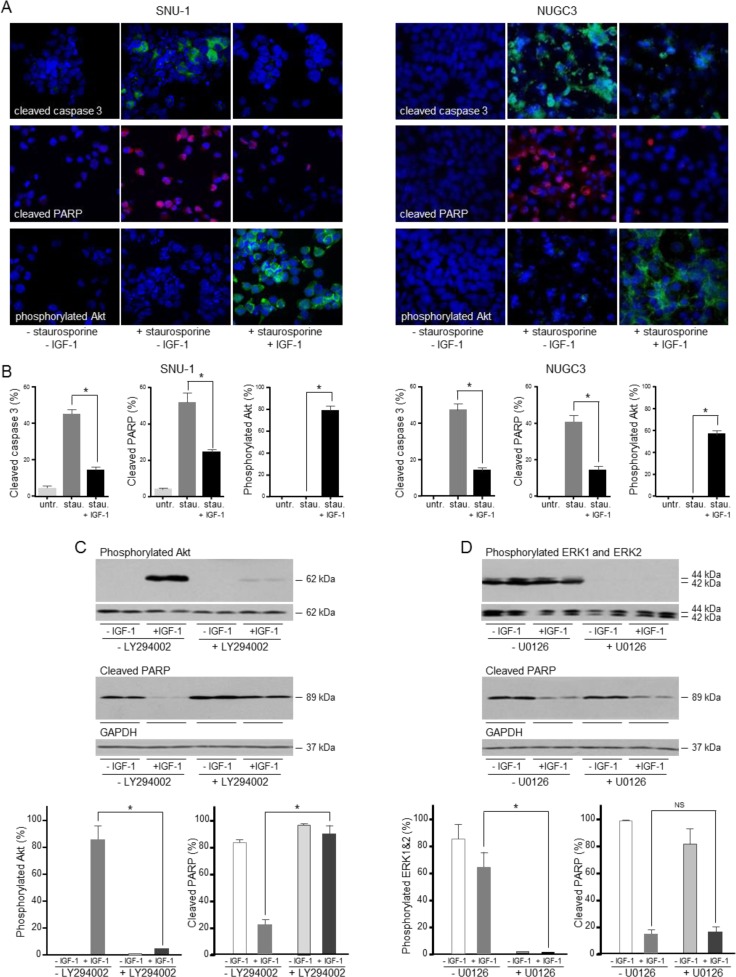
Figure 4. IGF protects gastric cancer cells from caspase-dependent cell death via the PI3-kinase/Akt pathway.
SNU-1 and NUGC3 cells were treated for 4 and 24 h, respectively with staurosporine (stau.) in the absence and presence of 50 ngml−1 IGF-1. Cells were fixed and incubated with antibodies against cleaved caspase 3, cleaved PARP and phosphorylated Akt A. and B. Nuclei were identified with the DAPI DNA dye. The proportion of cells with detectable cleaved caspase 3, cleaved PARP and phosphorylated Akt is shown as means ± SEM. Asterisks indicate differences that are statistically significant (One-way ANOVA; SNU-1, cleaved caspase 3, p < 0.0001; cleaved PARP, p = 0.0002; phosphorylated Akt, p < 0.0001; NUGC3, cleaved caspase 3, p < 0.0001; cleaved PARP, p < 0.0001; phosphorylated Akt, p < 0.0001). SNU-1 cells were treated with staurosporine in the absence and presence of 50 ngml−1 IGF-1 and 20 μM LY294002 or 6 μM U0126 inhibitor, lysed and cleaved PARP, phosphorylated Akt, ERK1 and ERK2 were measured and corrected for the expression of GAPDH or total corresponding protein C. Asterisks indicate phosphorylated protein levels that are significantly lower in the presence of an inhibitor than in its absence (Two-way ANOVA; phosphorylated Akt, p < 0.0001; phosphorylated ERK1 and ERK2, p = 0.0006) or cleaved PARP levels that are statistically significantly higher in the presence of an inhibitor (Two-way ANOVA; for LY294002 inhibitor, p = 0.0001. NS indicates values that are not significantly different.

