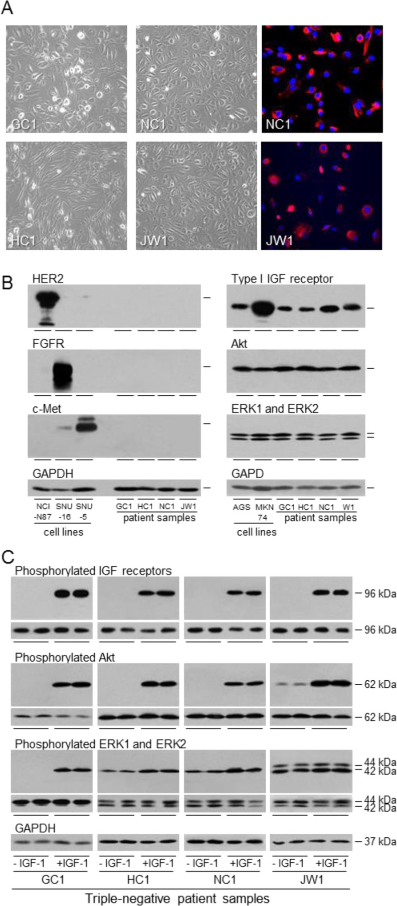
Figure 6. Expression and activation of the IGF signal transduction pathway in patient samples.
GC1, HC1, NC1 and JW1 cells were grown to 70% confluence and photographed or fixed and incubated with fluorescently-labelled antibody against epithelial cytokeratins A. Expression of HER2, FGFR2, c-Met, type I IGF receptor, Akt, ERK1 and ERK2 were analyzed by western transfer as described in the legend to Figure 1 B. GC1, HC1, NC1 and JW1 cells were withdrawn for two days and stimulated with 50 ngml−1 IGF-1 for 15 min. Phosphorylation of IGF receptors, Akt, ERK1 and ERK2 was analyzed by western transfer as described in the legend to Figure 2A.

