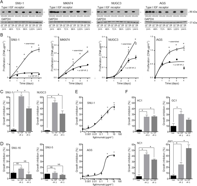
Figure 9. Importance of the type I IGF receptor in the growth of triple-negative gastric cancer cells and patient samples.
SNU-1, MKN74, NUGC3 and AGS cells were transfected with scrambled oligonucleotide (scr.) or si IGF-IR 2 (siR), cultured in DMEM and 10% FCS, lysed and type I IGF receptor and GAPDH A. or DNA content measured B. Asterisks indicate times at which there were significantly fewer cells after transfection with si IGF-IR 2 than with scrambled oligonucleotide (Two-way ANOVA; SNU-1, p < 0.0001; MKN74, p < 0.0001; NUGC3, p < 0.0001; AGS, p < 0.0001). SNU-1 and NUGC3 C., and SNU-5 and SNU-16 cells D. were transfected with scrambled oligonucleotides, si IGF-IR 2 or si IGF-IR 3 and cultured for 4 days, lysed and their DNA content measured. Asterisks indicate significant growth inhibition after reduction in receptor expression (One-way ANOVA; p < 0.001). NS indicates values that are not significantly different. SNU-1 and AGS cells were cultured in the presence of the indicated concentrations of the IGF inhibitory antibody figitumumab for 4 days. Cells were lysed and their DNA content measured. Asterisks indicate significant growth inhibition in the presence of figitumumab (One-way ANOVA; p < 0.001). GC1, HC1, NC1 and JW1 cells were transfected with scrambled oligonucleotides, si IGF-IR 2 or si IGF-IR 3 and cultured for 7 days in DMEM and 20% FCS E. Cells were lysed and their DNA content measured. Asterisks indicate significant growth inhibition after reduction in receptor expression (One-way ANOVA; HC1, p < 0.001; GC1, p= 0.0322; NC1, p < 0.0001; JW1, p = 0.0006).

