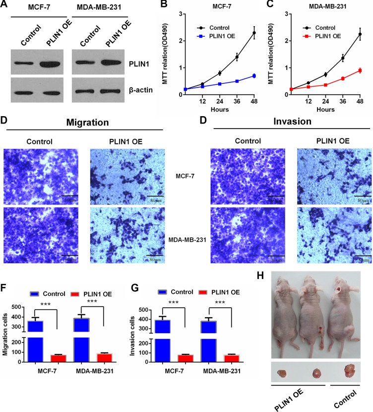
Figure 5. Effects of PLIN1 on human breast cancer cell proliferation, migration and invasion.
(A) Western blot assay shows the PLIN1 expression levels after transfection with the PLIN1-expressing recombinant plasmids in MCF-7 and MDA-MB-231 cells. (B–C) Cell proliferation analysis by MTT assays for MCF-7 (B) and MDA-MB-231 cells (C) with or without exogenous PLIN1. (D–G) Transwell assays show the effects of PLIN1 on MCF-7 and MDA-MB-231 breast cancer cell migration and invasion. Representative micrographs and statistical data exhibit the effects of PLIN1 on cell migration (D and F) and invasion (E and G). The data are presented as the mean values ± SD. The Two-tailed Student's t-test was used. *p < 0.05, **p < 0.01 and ***p < 0.001. (H) Representative pictures from a total of 6 tested and 6 control mice, showing tumorigenesis of hind limbs isolated from nude mice three weeks after injection of cells stably expressing PLIN1 or control cells.

