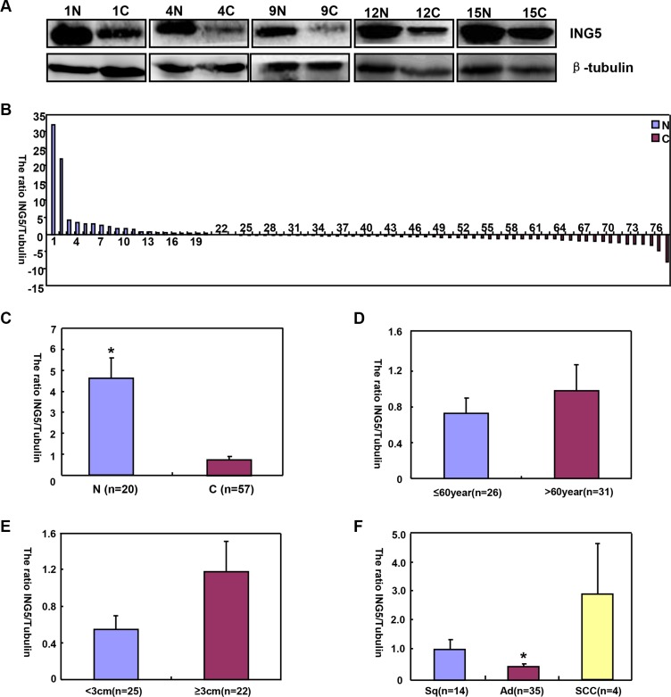
Figure 5. The correlation of ING5 protein expression with clinicopathological features of lung cancers.
Tissue lysate was loaded and probed with anti-ING5 antibody with β-tubulin as an internal control in normal tissue (N, n = 20) and lung cancer (C, n = 57) by Western blot. The densitometric analysis showed higher ING5 expression in normal tissue than in lung cancer (A–C, p < 0.05). ING5 expression was not related to age or tumor size (D and E), p > 0.05). ING5 was hypoexpressed in adenocarcinoma (Ad), in comparison to squamous cell carcinoma and small cell carcinoma (F, p < 0.05). The data is expressed as mean ± standard error; *p < 0.05.

