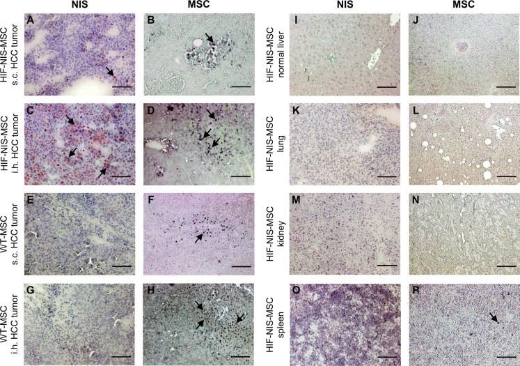
Figure 3. MSC recruitment and hypoxia-induced NIS expression were higher in intrahepatic compared to subcutaneous HCC tumors.
Compared to subcutaneous (s.c.) tumors (A), higher NIS-specific immunoreactivity was detected in intrahepatic (i.h.) HuH7 tumors (C). This correlated well with tumoral HIF-NIS-MSC recruitment (B, D). In mice injected with WT-MSCs, no NIS expression (E, G) was detected, though MSCs were recruited (F, H). Non-target organs showed neither MSC recruitment nor NIS expression (I–N), except for the spleen where no NIS staining (O) but positive MSC staining (P) were observed. One representative image is shown each. Scale bar = 100 μm.

