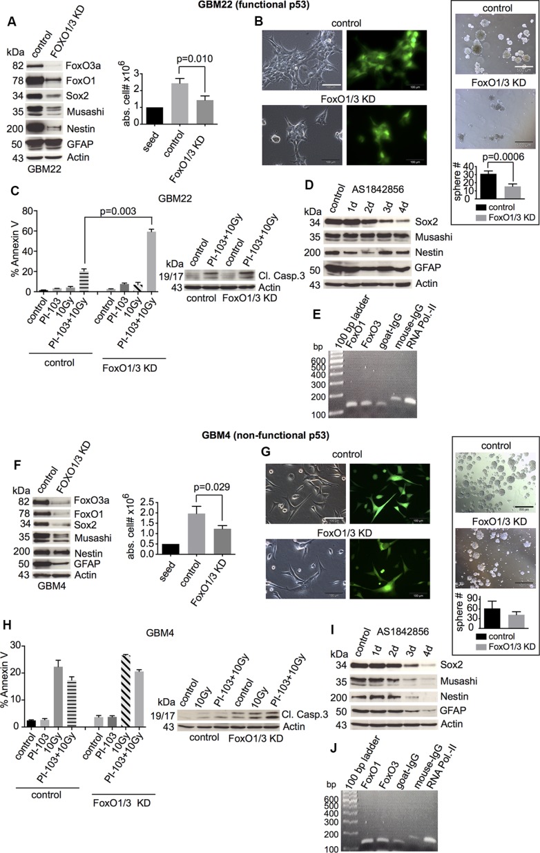
Figure 5. Effects of the combined FoxO1/3 knockdown on stemness and survival of GBM-SCs depending on p53 functionality.
(A and F, left) Western blot analyses of control shRNA- or FoxO1/3 shRNA-transduced GBM-SCs assessing the expression of FoxO proteins, stemness markers, and the differentiation marker GFAP (n = 3 experiments; for statistical analysis, see Figure S4). (A and F, right) Absolute cell numbers after 3 or 4 days of incubation in CSC medium. (B and G, left) Confirmation of the transduction efficiency by GFP expression analysis. The bar represents 100 μm. (B and G, right) Sphere-forming capacity after 3 days of incubation in CSC medium. The bar represents 500 μm. (C and H) Assessment of cleaved caspase 3 (1 of 2 experiments is shown, each with similar results) and of apoptosis by annexin V staining. Cells were treated with 0.5 μM PI-103 for 1 h and then irradiated with 10 Gy. The analyses were performed after 2 (GBM22) or 4 days (GBM4). (D and I) Western blot analyses of stemness markers and GFAP after incubation with 10 μM of the FoxO inhibitor AS1842856 (1 of 2 experiments is shown, each with similar results). (E and J) ChIP assay: DNA from GBM-SCs co-immunoprecipitated with anti-FoxO1, anti-FoxO3, or control antibody (goat IgG) was amplified by PCR using primers specific for the sox2 regulatory regions. Precipitation with anti-RNA polymerase II and the respective mouse IgG control antibody was performed to validate the assay (1 of 2 experiments is shown, each with similar results). Data for cell and sphere numbers and apoptosis in (A)–(C) and (F)–(H) represent means ± SD from 3 independent experiments. KD, knockdown.

