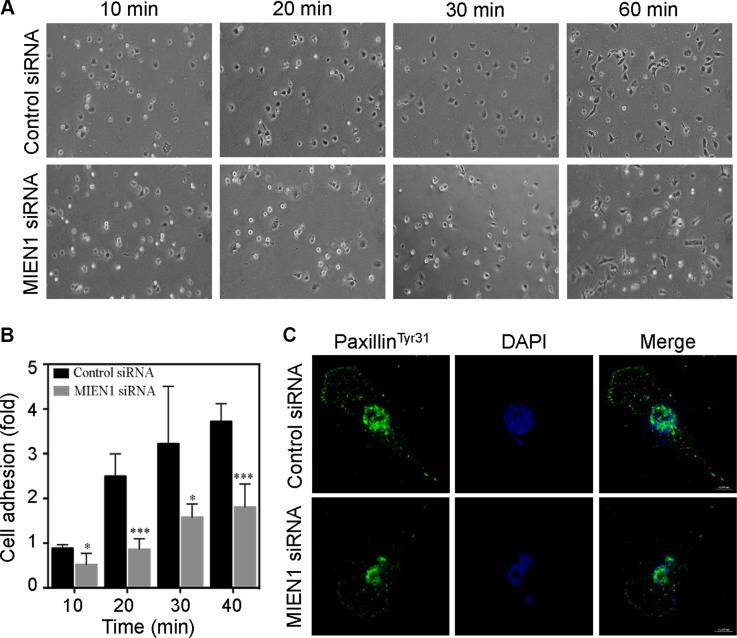
Figure 3. Depletion of MIEN1 affects focal complex turnover and cell adhesion.
(A) Phase imaging of control siRNA treated MDA-MD-231 cells plated on fibronectin-coated coverslips showed enhanced spreading and adhesion compared to MIEN1 knockdown cells. At the times indicated, phase-contrast micrographs were taken with a Nikon inverted microscope equipped with a 10X objective lens. (B) The data represent quantification of adherent cells from 5 random sites. The experiment was repeated twice. (*p < 0.05; ***p < 0.001 vs same time-point control). (C) MDA-MD-231 cells were cultured on fibronectin-coated coverslips and transfected with control or MIEN1 siRNA for 72 h. Coverslips were fixed and stained with paxillinTyr31 (green). Nuclei were stained with DAPI (blue).

