
Figure 4. HBxΔ31 transcriptionally represses the RhoGDIα and CAPZB expressions through enhancing MAZ binding to the promoter.
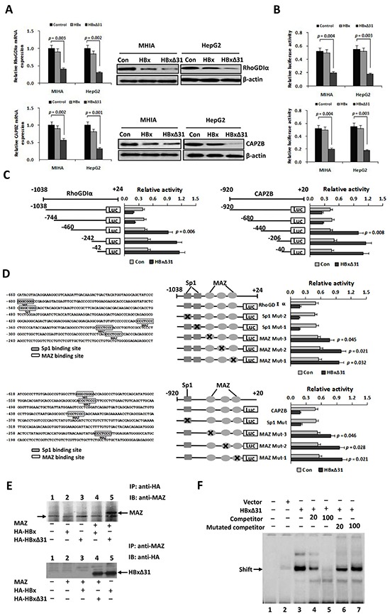
A. Real-time PCR and Western blot analysis showing that HBxΔ31 repressed RhoGDIα and CAPZB expressions in MHIA and HepG2 cells. B. Dual luciferase report assay showing that HBxΔ31 repressed the RhoGDIα and CAPZB promoters. C. The sequence analysis demonstrated that the HBxΔ31 repressive element located between nt.-744 to −460 of the RhoGDIα promoter, while between nt. −680 to −440 of the CAPZB promoter. On the left panel, the schematic representation of the reporter gene constructs is shown; on the right panel, bars represent the relative promoter activity in each of the transfected cells. D. On the left panel, analysis of the cis-regulatory elements between nt. −744 to −460 in the RhoGDIα promoter revealed 2 Sp1 binding sites (GGGCGGG) and 3 MAZ binding sites (CCCTCCC), whereas between nt. −680 to −440 in the CAPZB promoter exhibited 1 Sp1 binding sites and 3 MAZ binding sites. On the right panel, MAZ sites in the RhoGDIα or CAPZB promoter was essential for HBxΔ31-induced RhoGDIα or CAPZB trans-suppression. Mutations in the Sp1 sites had no effect on the RhoGDIα and CAPZB promoter activities, whereas mutations in the MAZ sites significantly enhanced their promoter activities regulated by HBxΔ31. E. Coimmunoprecipitation assay showed a direct binding of HBxΔ31, but not full-length HBx, to MAZ in HCC cells. The arrow marks the location of a background band. F. EMSA assay confirmed enhancement of the DNA binding activity of MAZ to the RhoGDIα promoter by HBxΔ31.