
Figure 5. Cilengitide (CIL) inhibits pulmonary metastasis in vivo .
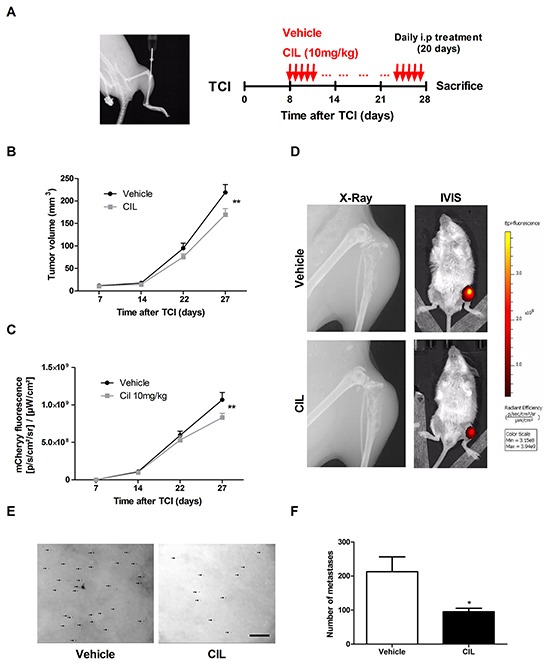
A. X-Ray image of the site of intratibial injection of 143-B/mCherry/LacZ cells taken at the time of the tumor cell injection (TCI) on Day 0 (left panel). Protocol for the treatment of tumor bearing mice with vehicle (control) or CIL (10 mg/kg body weight) (right panel). B. Primary tumor growth over time monitored by caliper measurements of the tumor volume at indicated time points in mice treated with vehicle or with CIL. C. mCherry fluorescence indicating primary tumor growth over time in the same vehicle- or CIL-treated animals. D. X-Ray images of tumor bearing legs and IVIS images of tumor bearing animals taken at the end of the experiment from representative mice treated with vehicle or with CIL. E. Representative pictures and F. quantification of X-gal stained pulmonary metastases (arrows) on lung mounts collected and prepared as described in the Materials and Methods from vehicle or CIL treated mice at sacrifice 28 days after TCI. Scale bar, 250 μm. Values in B, C and F are the mean ± SEM of the data collected in 12 vehicle- and 11 CIL-treated mice; *, P < 0.05; **, P < 0.01.