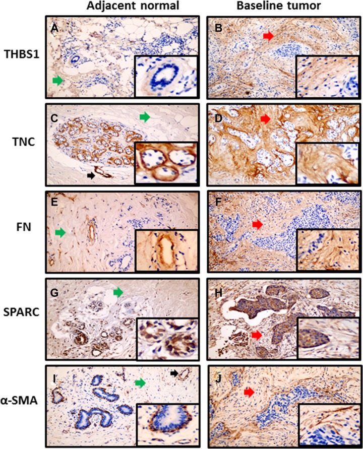
Figure 1. Immunoreactivity of stromal proteins in baseline tumor with matched adjacent normal tissue.
Magnification 100× and 400× (inserted pictures). (A, C, E, G, and I) Weak stromal proteins expression in the stroma area (green arrows) of adjacent normal tissue. Black arrows in panel C and panel I showed positive staining in blood vessels. (B, D, F, H and J) Moderate to strong stromal proteins expression in the stroma area (red arrows) of matched baseline tumor.

