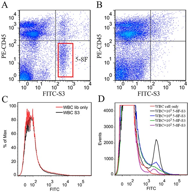
Figure 6. Recognition of the target cells in real peripheral blood samples by S3.

A. 105 NPC 5-8F cells dispersed in 1mL of peripheral blood from healthy volunteer. B. 105 A549 cells dispersed in 1mL of peripheral blood from healthy volunteer. PE-labeled anti-CD45 monoclonal antibody was used to recognize the white blood cells (WBCs). C. Flow cytometric analysis of aptamer S3 with WBCs. D. Flow cytometric analysis for the binding of the FITC-labeled aptamer S3 with 105, 104, 103 and 102 5-8F cells dispersed in 1 mL of peripheral blood.
