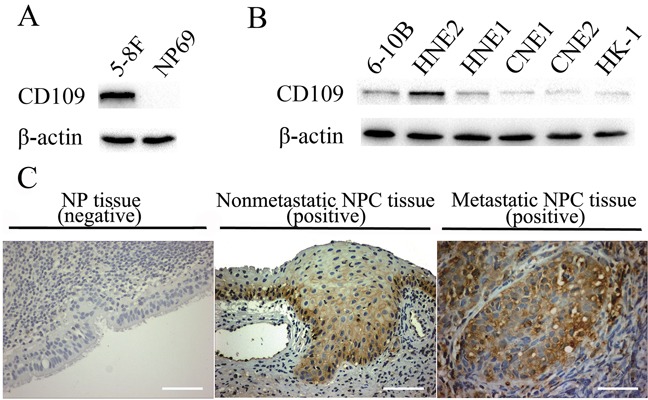
Figure 9. Expression of CD109 in different cell lines and immunostaining of CD109 in clinical NPC and NP tissues.

A. The expression of CD109 in 5-8F and NP69 cells. B. The expression of CD109 in 6 other NPC cell lines. CD109 was expressed in all the tested NPC cell lines, but not detectable normal NP69 cells.C. Immunostaining of CD109 in clinical NPC and NP tissues. CD109 was positively expressed in both nonmetastatic and metastatic NPC tissues, but it was not detectable or fairly low in NP control tissues. (Scale bar, 100 μm)
