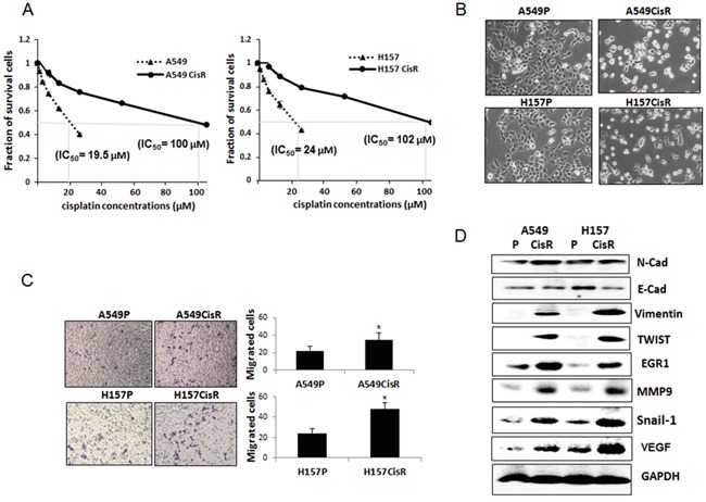
Figure 1. EMT and metastatic potential were increased in cisplatin-resistant NSCLC cells compared to parental cells.

A. Cytotoxicity test of A549P/H157CisR and H157P/H157CisR cells against cisplatin treatment. Cisplatin-resistant cells were obtained by continuous treatment of cells with increasing dose of cisplatin for 6 months. Cisplatin cytotoxicities of A549P/H157CisR and H157P/H157CisR cells were analyzed in the presence of various concentrations of cisplatin in MTT assay. B. Morphology test. A549P/H157CisR and H157P/H157CisR cells (1 × 10³) were seeded and morphology was observed under microscope. C. Migration test. Cells (A549P/A549CisR and H157P/H157CisR, 1 × 10⁴) were placed in upper chamber of transwell plates (8 μm pore) and migrated cells to lower chamber were counted under microscope after crystal violet staining at the end of 24 hours of incubation. Quantitation shown on right. D. Western blot analysis showing an increase in EMT/metastasis markers in cisplatin-resistant cells compare to parental cells. Cell extracts were obtained from A549P/A549CisR and H157P/H157CisR cells and Western blot analyses were performed using antibodies against indicated molecules. *p<0.05, **p<0.01.
