
Figure 4. In vivo mice studies.
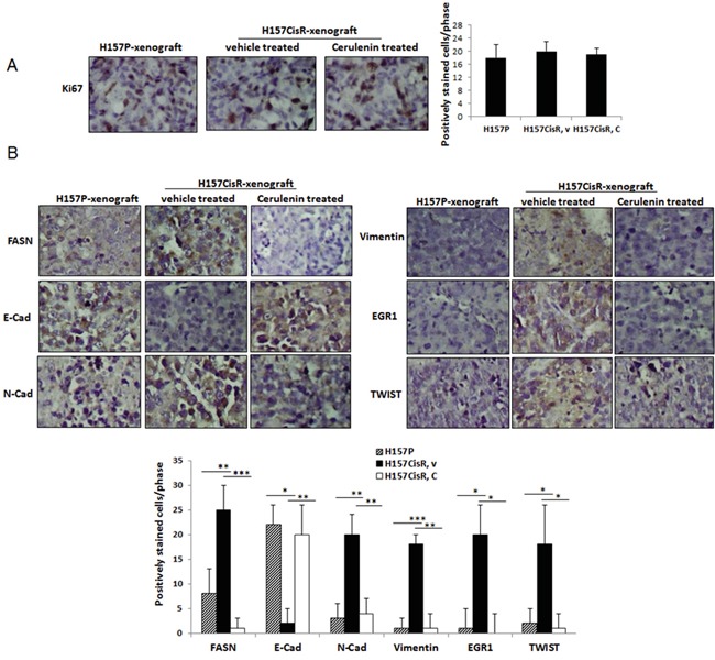
A, B. IHC staining of tumor tissues using antibodies of Ki67 (A) and EMT/metastasis markers (B). Mice (H157CisR cell-derived xenograft) were sacrificed 3 days at the completion of cerulenin (or vehicle) treatment (H157P cell-derived xenografts did not receive treatment) and tumor tissues were obtained. The processed tumor tissues were cut into 5 μm, and stained with antibodies of FASN and EMT/metastasis markers. For Ki67 staining, tissues were subjected to antigen retrieval before staining. Magnification, x 100. Quantitation shown below. *p<0.05, **p<0.01, ***p<0.001.