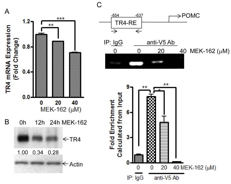
Figure 4. MEK-162 treatment reduces TR4 expression and inhibits binding to POMC promoter.
A., Q-PCR was used to quantitate TR4 mRNA in murine corticotroph tumor AtT20 cells following MEK-162 treatment (20 μM and 40 μM for 24 h). B., TR4 protein expression was detected by Western Blotting in murine corticotroph tumor AtT20 cells following 40 μM MEK-162 treatments for 12 h and 24 h, and quantified using densitometric analyses of the TR4 protein bands vs. the individual loading controls. C., AtT20 cells were transiently transfected with V5-TR4 plasmid. 24 h later, cells were treated with 20 μM and 40 μM MEK for 24 h, after which ChIP assay was performed to measure TR4 binding to the POMC promoter (−854bp~ −637bp). Each bar indicates the mean ± standard error of triplicate tests. * p < 0.05; ** p < 0.01, *** p < 0.005. Data shown were representative of at least three independently repeated experiments.

