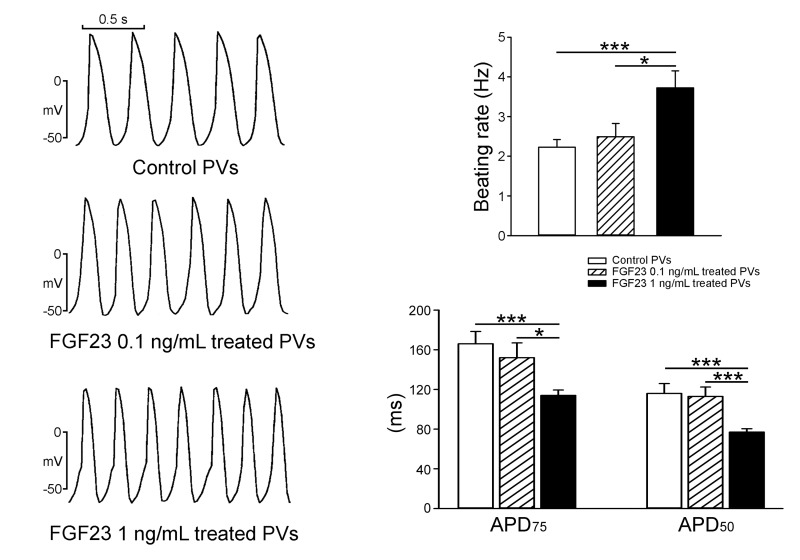
Figure 1. Spontaneous beating rates and action potential duration (APD) of pulmonary vein (PV) cardiomyocytes in the control and different concentrations of FGF23.
An example and average data show that FGF23 (1 ng/mL)-treated PV cardiomyocytes (n = 10) had more-rapid beating rates and shorter APD than control (n = 10) or FGF23 (0.1 ng/mL)-treated PV cardiomyocytes (n = 10). * p < 0.05, *** p < 0.005 vs. the control.

