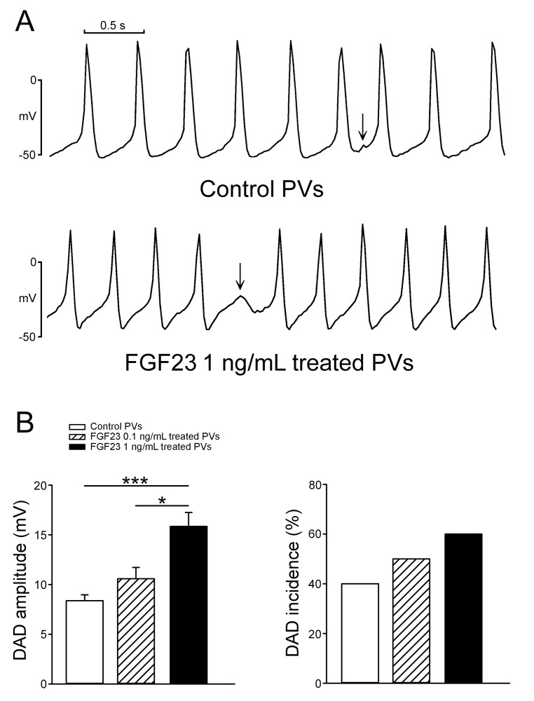
Figure 2. The incidence and amplitude of delayed afterdepolarizations (DADs) in control and FGF23-treated PV cardiomyocytes.
A. Tracings show that a FGF23 (1 ng/mL)-treated PV cardiomyocyte had a larger amplitude of DAD (arrow) than a control PV cardiomyocyte. B. In the cells with DADs, and FGF23 (1 ng/mL, n = 6)-treated PV cardiomyocytes had larger amplitude of DAD than control (n = 4) and FGF23 (0.1 ng/mL, n = 5)-treated PV cardiomyocytes. However, the incidence of DAD was not significantly different in control (n = 10), and FGF23 (0.1 ng/mL, n = 10) and FGF23 (1 ng/mL, n = 10)-treated PV cardiomyocytes.* p < 0.05, *** p < 0.005.

