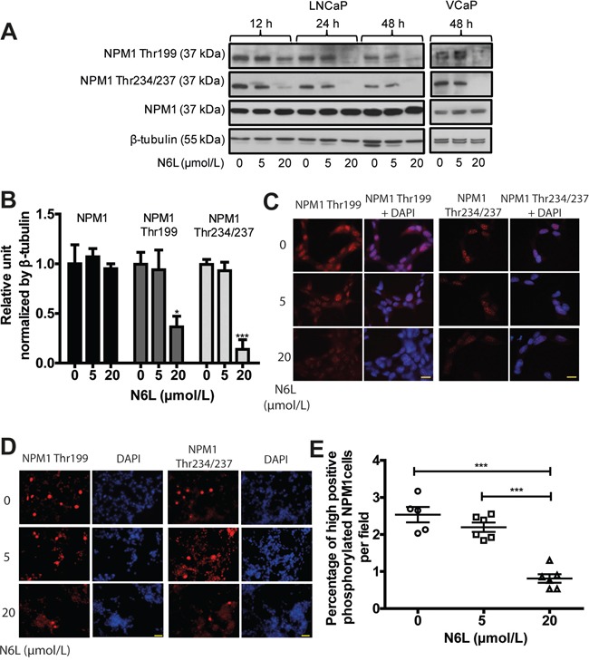
Figure 2. N6L reduced NPM1 phosphorylation on Thr199 and Thr234/237.

LNCaP or VCaP cells were treated for 12, 24 or 48 hours with or not 5 or 20 μmol/L N6L. A. Protein expressions analyzed by Western blot. B. Densitometry quantification (ImageJ software) of protein expressions of LNCaP cells treated or not for 48 hours with N6L showed by protein/β-tubulin ratio density. Means ± sd (n = 3), * p < 0.05 and *** p < 0.001. C and D. LNCaP cells, treated or not with N6L for 48 hours, were fixed with methanol, immunostained (Thr199 and Thr234/237 NPM1) and analyzed by spinning disk fluorescence microscopy at high (C: scale bar, 5 μm) or low magnification (D: scale bar, 20 μm). Nuclei were stained with DAPI. E. Quantification of cells displaying strong staining of phosphorylated Thr199 or Thr234/237 NPM1. Data are expressed as the percentage of strong positive cells from 5 pictures per condition. Means ± sd (n = 5 for control and 6 for treated cells), *** p < 0,001.
