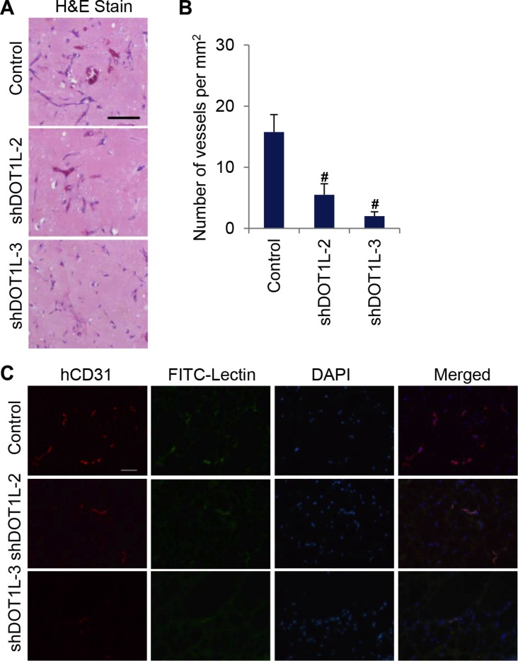
Figure 3. Depletion of DOT1L inhibits angiogenesis in vivo.
Matrigel plugs containing HUVECs expressing DOT1L shRNA2, shRNA3 or control shRNA, were subcutaneously implanted in SCID mice in vivo. Eight days later, hematoxylin and eosin staining was performed, and matrigel plugs were stained with anti-hCD31 and FITC-Lectin to analyze vascularization. (A) Representative hematoxylin and eosin–stained plug sections. Scale bar, 55 μm. (B) Quantification of microvessel density determined by the number of vessels per square milimeter. Data are mean ± SD for n = 4; #P < 0.01 vs. control (Student's t test). (C) Fluorescent confocal microscopic images of frozen sections of matrigel plugs. Red, hCD31. Green, FITC-Lectin. Blue, DAPI. Scale bar, 100 μm.

