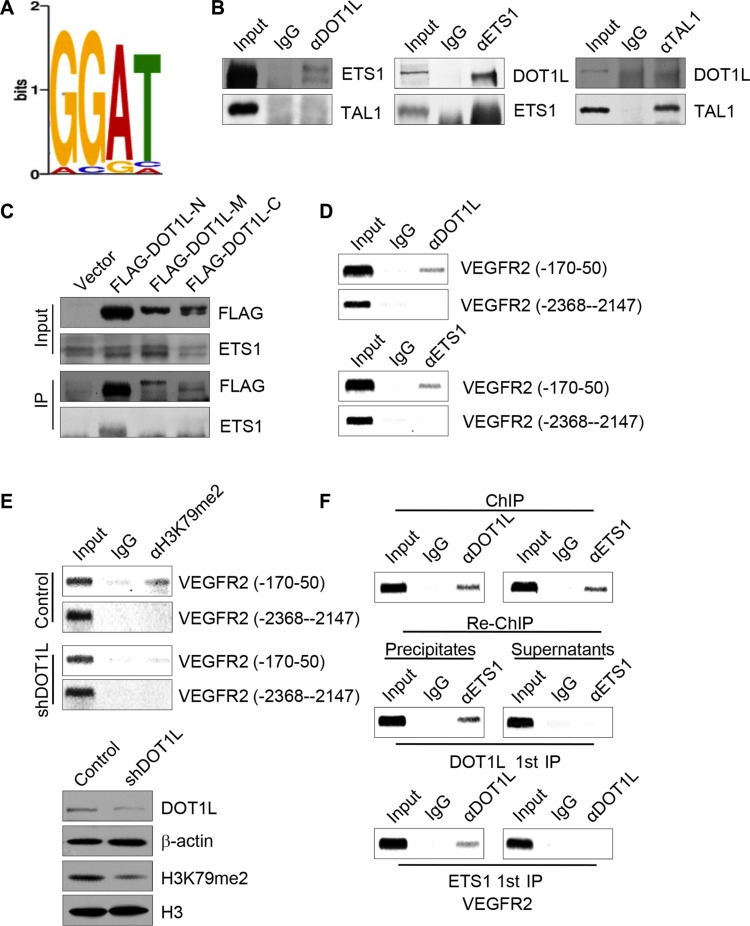
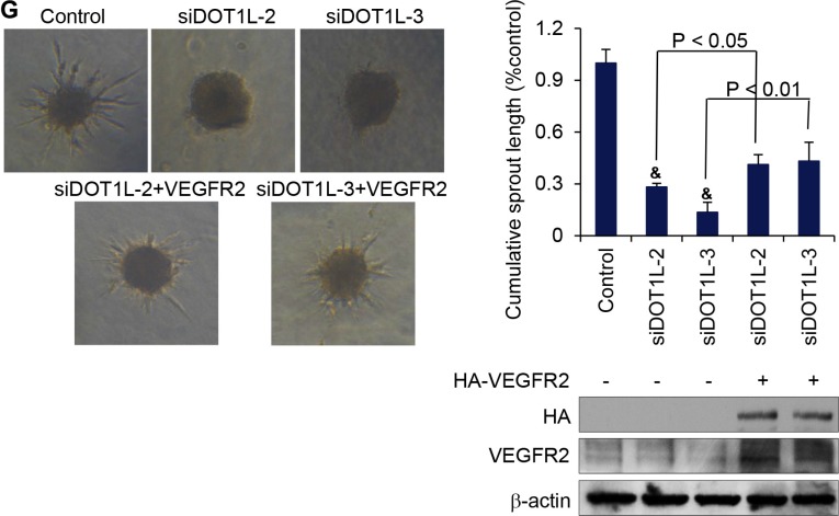
Figure 6. DOT1L cooperates with ETS-1 to regulate the transcription of VEGFR2.
(A) H3K79me2 enriched motifs was analyzed using MEME suite. (B) Endogeous interaction of DOT1L with ETS-1. Immunoprecipitation assays were performed with antibodies against indicated proteins followed by immunoblotting with antibodies against indicated proteins. (C) Identification of domains responsible for the interaction between DOT1L and ETS-1. Whole-cell lysates from HUVECs constantly expressing FLAG-fused DOT1L truncation mutants were prepared and immunoprecipitation was performed with anti-FLAG, followed by immunoblotting with antibodies against ETS-1. (D) The recruitment of DOT1L and ETS-1 on the downstream of VEGFR2 promoter. ChIP assays were performed in HUVECs using the indicated antibodies and primer pairs on VEGFR2 promoter and gene body. (E) DOT1L depletion led to decreased H3K79me2 occupation on VEGFR2 gene body. ChIP assays were performed in HUVECs expressing DOT1L shRNA or control shRNA using antibodies against H3K79me2 and indicated primers. Western blotting was also performed with anti-DOT1L, anti-β-actin, anti-H3K79me2 or anti-H3. (F) DOT1L and ETS-1 exist in the same complex on the downstream of VEGFR2 promoter. ChIP and Re-ChIP assays were performed with indicated antibodies. (G) VEGFR2 was overexpressed in DOT1L depleted-HUVECs, and angiogenic sprouting was compared in the spheroid model. Representative spheroids are shown. Endothelial sprouting capacity is given as cumulative sprout length per spheroid. Data are mean ± SD for n = 3; &P < 0.05 vs. control (Student's t test).


