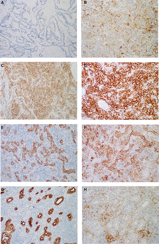
Figure 5. Representative images of PD-L1 immunostaining.

PD-L1 was immunostained on the membrane and/or in the cytoplasm of tumor cells with variable intensities: (A–D) absent in the conventional IHCC (score 0), weak in LELCC (score 1), moderate in LELCC (score 2), strong in LELCC (score 3). In most cases, PD-L1 was expressed in tumor cells, which were highlighted by (E) AE1/AE3, and (F) tumor-infiltrated immune cells. (H) In 3 cases, PD-L1 was immunostained mainly in tumor-infiltrated immune cells, (G) with little staining in tumor cells, which were highlighted by AE1/AE3. Immunohistochemistry, ×200.
