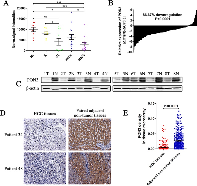
Figure 1. PON3 is frequently decreased in HCC.
(A) Normalized signal intensities of PON3 in normal liver (NL), chronic inflammatory liver (IL), cirrhotic liver (CL), early-stage HCC (eHCC), and advanced-stage (aHCC) tissues in a microarray (GSE54238). (B) PON3 mRNA level in 135 paired HCC and the adjacent non-tumor tissues were evaluated by qRT-PCR. (C) Western-blots showing PON3 protein level in tumor tissues (T) and the paired adjacent non-tumor tissues (N) from eight HCC patients. (D) Two representative cases of PON3 IHC staining in HCC and adjacent non-tumor tissue pairs in tissue microarray. (E) Relative IHC staining of PON3 in paired HCC and adjacent non-tumor tissue samples (n = 286). Statistical significance was determined by student's t-tests. *p < 0.05; **p < 0.01; ***p < 0.0001 compared with respective controls. Data are shown as mean ± SD.

