
Figure 4. Down-regulation of PD-1 induced by CpG is dependent on IL-12.
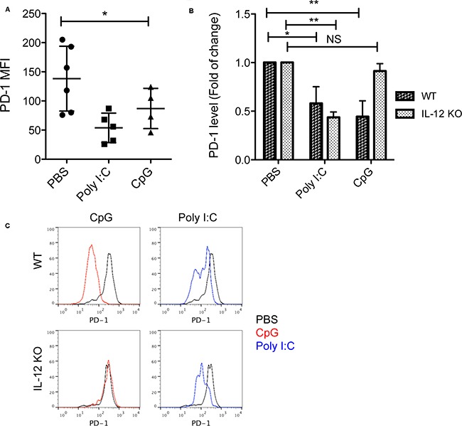
A. PD-1 levels (MFI) among tumor-reactive CD11ahigh CD8+ T cells isolated from tumors one day after last treatments with CpG, Poly I:C or PBS. Data show mean ± s.d. of 4-6 mice per group, *P<0.05 compared with PBS groups (One-way ANOVA). B. Fold of Changes of PD-1 levels in tumor-reactive CD8+ T cells isolated from tumors treated with CpG or Poly I:C in WT mice and IL-12 KO mice (n=4). *P<0.05, **P<0.01 compared with PBS groups in WT or IL-12 KO mice (One-way ANOVA). NS: non-significant. C. Representative histograms show PD-1 expression by CD11ahigh CD8+ T cells in tumor tissues following treatment with CpG or Poly I:C in WT or IL-12 KO mice. Black lines show the baseline levels of PD-1 in PBS treated groups.