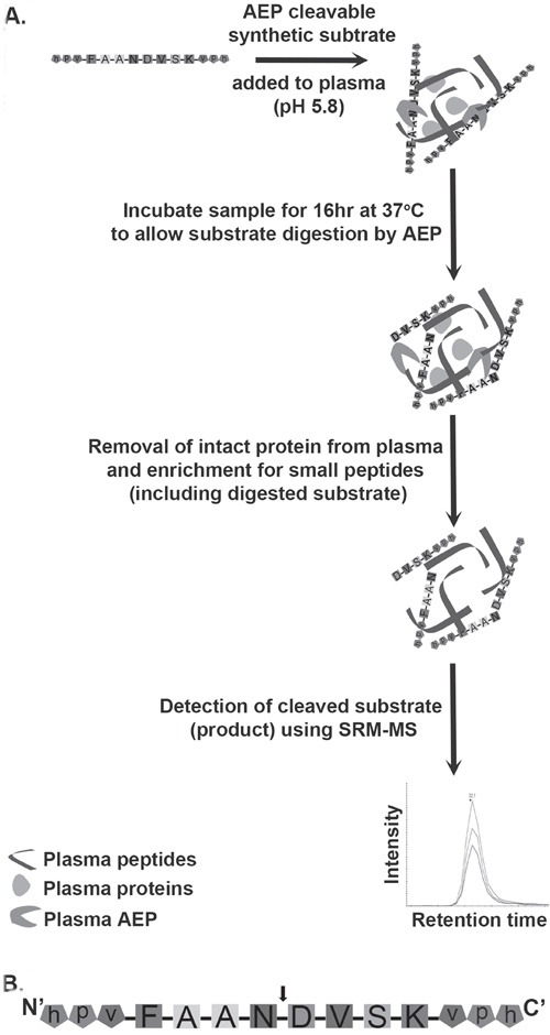
Figure 1. Schematic diagram of the workflow involved in assaying AEP activity in plasma samples.

A. The workflow to measure AEP activity in plasma. AEP cleavable synthetic substrate was added to plasma at an acidic pH (pH 5.8) followed by an incubation at 37°C for 16 hr. This incubation allows endogenous plasma AEP to interact with synthetic substrate and cleaves substrate into specific products. Removal of intact proteins from plasma using acetonitrile (ACN) precipitation gives enrichment of peptides including products formed by digestion of synthetic substrate by AEP. The cleaved form of substrate is detected by mass spectrometer following SRM based approach. B. AEP cleavable synthetic substrate peptide (FAANDVSK) was designed and synthesized with a cleavage site for AEP at the C-terminal of asparagine (N) residue. The arrow head represents the AEP cleavage site. Both the N and C terminals of the substrate were protected by using a D-amino acid capping (represented by lower case).
