
Fig. 1. Wearable/disposable sweat monitoring device and microneedle-based transdermal drug delivery module.
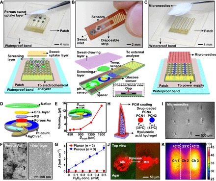
(A) Optical camera image (top; dotted line, edges of the patch) and schematic (bottom) of the wearable sweat monitoring patch. A porous sweat-uptake layer is placed on a Nafion layer and sensors. (B) Optical camera image (top) and schematic (bottom) of the disposable sweat monitoring strip. (C) Optical camera image (top; dotted line, edges of the patch) and schematic (bottom) of the transdermal drug delivery device. Replacement-type microneedles are assembled on a three-channel thermal actuator. (D) Schematic drawing of the glucose sensor in an exploded view. PB, prussian blue. (E) Minimum volume of the artificial sweat required for sensing with different sizes of the glucose sensor. (F) Scanning electron microscope (SEM) images before (left) and after (right) immobilization of the enzyme (enz.) on the porous gold electrode. (G) Comparison of the H2O2 sensitivity in the planar and porous gold electrode deposited with Prussian blue at different H2O2 concentrations. (H) Schematic of the drug-loaded microneedles. The right inset describes details of different PCNs. (I) SEM image of the microneedles. (J) Confocal microscope images of the released dye from microneedles (MN) (top view) into the 4% agarose gel (green, agar; red, dye). (K) Infrared (IR) camera image of the three-channel (ch) thermal actuator.