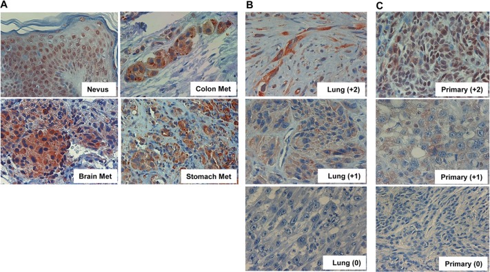
Figure 1. Heparanase expression in metastatic melanoma.
A. Melanoma metastases were subjected to immunostaining applying anti-heparanase antibody as described under ‘Materials and Methods’. Shown is representative staining of heparanase in brain, colon, and stomach melanoma metastases. Heparanase immunostaining in non-malignant nevi is also included as a reference for non-malignant lesion. Note nuclear localization of heparanase in non-malignant skin tissue, compared with diffused cytoplasmic distribution in melanoma metastases. Representative lung metastases exhibiting strong (+2), weak (+1) or no staining (0) of heparanase are shown in (B). Immunostaining of heparanase in non-matched primary melanomas exhibiting strong (+2), weak (+1) or no (0) staining is shown in (C). Original magnifications: x100.

