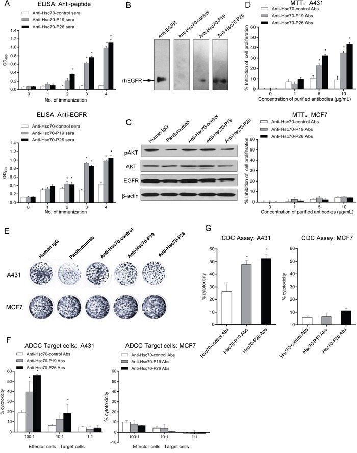
Figure 3. Antibody responses induced by mimotope immunization.

A. ELISA was performed to determine the anti-peptide (upper panel) and anti-EGFR (lower panel) antibody responses with GST fusion protein and rhEGFR-coated plates, respectively. Mice sera were diluted at 1:2000 and 1:100, respectively. B. Western blot analysis of anti-EGFR antibody from immunized mice. C. Western blot to determine the role of mimotope antibodies on EGFR-mediated signaling. D. MTT assays of A431 (upper panel) and MCF7 cells (lower panel) treated with purified mimotope antibodies. E. Colony formation assay of A431 and MCF7 cells treated with purified mimotope antibodies. F. ADCC activity of anti-mimotope antibodies. Human PBMC were used as effector cells. The effector to target A431 (left panel) or MCF7 cells (right panel) (E:T) ratio was 1:1, 10:1, and 100:1. Cytotoxicity was calculated by the formula: (experimental - effector spontaneous - target spontaneous) ×100%/(target maximum - target spontaneous). G. CDC activity of mice immunized with mimotopes. A431 (left panel) or MCF7 cells (right panel) were cultured with purified mimotope antibodies and human sera. The cytotoxicity was calculated. Data are shown as the mean (±SDs) of 3 independent experiments performed in triplicate. Statistically significant differences are indicated. *, P < 0.05.
