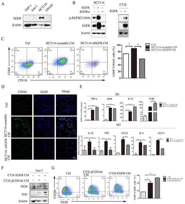
Figure 2. Inhibition of the EGFR signaling pathway in colon cancer cells prevents conditioned medium-induced M2-like macrophage polarization.

A. EGFR protein levels in THP-1, Ana-1, HCT116, and SW620 cells were detected by Western blot. B. HCT116 cells were cultured to 50% confluence and then transfected with human scramble siRNA, pCDNA6-EGFR WT plasmid, or EGFR siRNA. CT26 cells were cultured to 50% confluence and then transfected with human pCDNA6 vector or pCDNA6-EGFR WT plasmid for 48 h; the cells were then harvested for Western blots for EFGR. C. Percentages of CD68+/CD11b+ in THP-1 cells after 48 h of treatment with normal RPMI1640, HC116 scramble CM, or HCT116 siEGFR CM were detected by flow-cytometry. D. Immunofluorescent staining for CD206+ was measured in THP-1 cells after incubation with normal RPMI1640, HCT116 scramble CM, or HCT116 siEGFR CM. E. M1-related marker (TNF-α, iNOS, IL-12 and CCR7) and M2-related marker (IL-4, CCL17, CCL22, IL-10 and Arg1) mRNA levels were detected by q-PCR in THP-1 cells after incubation with normal RPMI1640, HCT116 scramble CM, or HCT116 siEGFR CM. Scale bars: 100 μm. F. Arg1 and iNOS protein levels in Ana-1 cells were detected by Western blot after incubation with CT26 pCDNA6 CM or CT26 EGFR CM. G. Percentages of F4/80+/CD206+ in Ana-1 cells after incubation with CT26 pCDNA6 CM or CT26 EGFR CM were detected by flow cytometry. Red arrows indicate CD206 expression in the cell membrane. Nuclei were counterstained with DAPI. Bars represent means ± SD (n = 3) for each treatment.*p < 0.05; **p < 0.01; ***p < 0.001.
