
Figure 3. EGFR knockout in colon cancer cells inhibits macrophage-induced promotion of xenograft tumor growth.
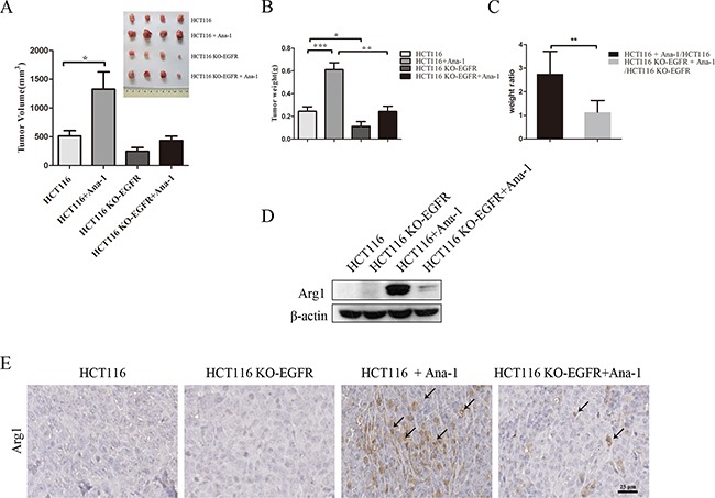
A. Mouse tumor volume quantification (bottom left). Images (top right) of tumors resulting from subcutaneous injection of HCT116 cells alone, HCT116 plus Ana-1 cells, HCT116 KO-EGFR cells alone, and HCT116 KO-EGFR plus Ana-1 cells (n=4) into Balb/c nude mice. After 18 days, the mice were sacrificed and tumors were excised. The experiment was repeated twice. B. Mouse tumor weights were measured. C. Weight ratio of mice receiving HCT116 plus Ana-1/HCT116 injections compared to HCT116 KO-EGFR plus Ana-1/HCT116 KO-EGFR injections. D. Arg1 protein levels were detected by Western blot in HCT116, HCT116 KO-EGFR, HCT116 plus Ana-1, and HCT116 KO-EGFR plus Ana-1 mouse tumors. E. Immunostaining for Arg1 in xenograft mouse tumor tissues. Scale bars: 50 μm. Bars represent means ± SD (n = 3) for each treatment. *p < 0.05; **p < 0.01.