
Figure 4. Curcumin directly targets TFEB for activation.
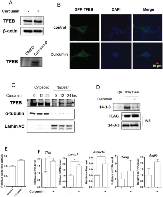
A. HCT116 cells were treated with 10 μM Curcumin for 12 hours and cell lysates were prepared followed by immunoblotting for TFEB and β-actin (up panel). HCT116 cells were labeled with Curcumin-probe (30 μM) for 4 hours and western blotting was performed to validate Curcumin-probe targeted TFEB (down panel). B. Enhanced TFEB nuclear translocation in response to Curcumin treatment (10 μM; 12 hours). Live imaging of GFP-TFEB (green) and DAPI (blue) in HCT116 cells showed an enrichment of the GFP-TFEB signal in the nuclear. Five fields containing 20 to 30 cells were analyzed for TFEB nuclear localization. Scale bar, 10 μm. C. HCT116 cells were treated with 10 μM Curcumin as indicated. Cytosolic and nuclear fraction from control and Curcumin-treated cells were probed for TFEB. The same membrane was then stripped and reprobed for α-tubulin or Lamin AC as loading control. D. HCT116 cells were transient transfected with the TFEB-3x Flag (kindly provided by Dr. A Ballabio) and then treated with 10 μM Curcumin for 12 hours. Cells were lysed and subjected to immunoprecipitation with anti-FLAG antibody followed by immunoblotting for 14-3-3. TFEB was also determined using anti-FLAG antibody. E. HCT116 cells were transiently transfected with the TFEB-luc reporter construct (kindly provided by Dr. A Ballabio). After 24 hours, the cells were treated with Curcumin (10 μM) for another 12 hours and the relative luciferase activity was measured. RLU refers to relative luciferase units. Error bars represent the standard deviation from two independent experiments. F. HCT116 cells were treated with Curcumin (10 μM) for 12 hours and cells were harvested for RNA extraction. Real-time PCR was performed to determine mRNA level changes in the known TFEB target genes, such as Lamp1, Atp6v1a, Uvrag and Atg9b. Gapdh was used as a loading control. All values are means ± SD at least 3 independent experiments. Student's t test, * P < 0.05.