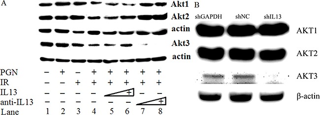
Figure 6. IL13 preferentially stimulated AKT3.

A. Western blot analysis of AKT1/2/3 expression in untreated, PGN alone-treated, radiation alone-treated, or radiation + PGN treated HCT116 cells. Lanes 5 and 6 reflect addition of 0.8 and 1.2 ng/mL IL13, respectively. The concentrations of IL13 neutralizing antibody in lanes 7 and 8 were 0.12 and 0.2 μg/mL, respectively. β-actin was used as a loading control. B. shIL13/shNC/shGAPDH plasmids were transfected into HCT116 cells, as indicated. AKT1/2/3 was detected by western blot after 48 h. β-actin was used as a loading control.
