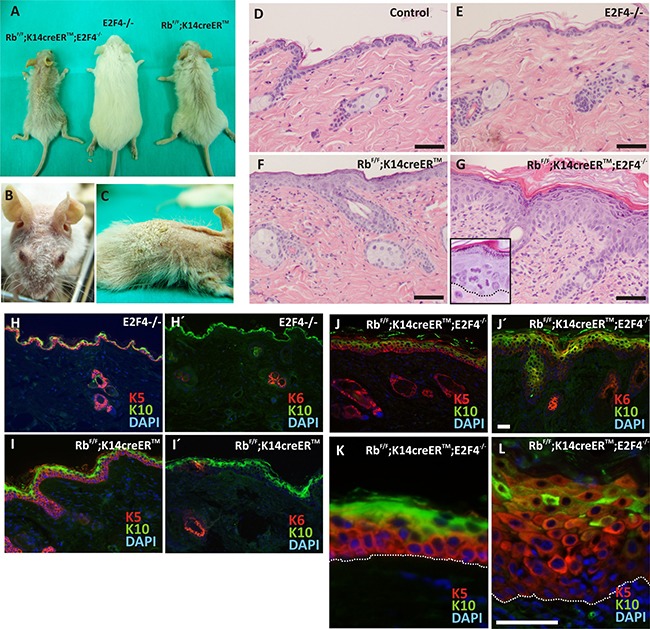
Figure 1. Phenotypic characterization of RbF/F;K14creERTM;E2F4−/− mice.

A. Gross appearance of the RbF/F;K14creERTM;E2F4−/−; E2F4−/− and RbF/F;K14creERTM mice 12 months after topical tamoxifen treatment. B, C. Macroscopic aspect of RbF/F;K14creERTM;E2F4−/− head and back respectively. D-G. H&E stained skin sections of back skin samples of Control (D); E2F4−/− (E); RbF/F;K14creERTM (F) and RbF/F;K14creERTM;E2F4−/− (G) mice. H-L. Representative double immunofluorescence of K5 or K6 (red) and K10 (green) of the quoted genotypes. Nuclei are stained in blue with DAPI. Bars = 100 μm (H-J Bars =50 μm).
