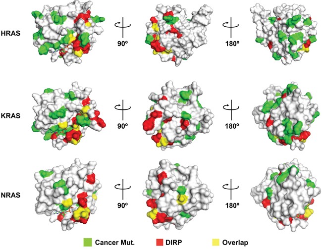
Figure 8. DIRP overlap with or are positioned near to residues frequently mutated in cancer.

Shown are the surface models for the 3D structures of HRAS (pdb# 1aa9), KRAS (pdb# 4epv) and NRAS (pdb# 3con; the only NRas 3D structure available in PDB, lacking residues 61-71), the three human RAS paralogs most frequently mutated in cancer. Mutations currently included in the TCGA catalog (Cancer Mut.) have been colored in green and DIRP in red. Overlapping positions (i.e., DIRP corresponding to residues mutated in cancer) are colored in yellow. For clarity, three rotating views are shown for each Ras protein.
