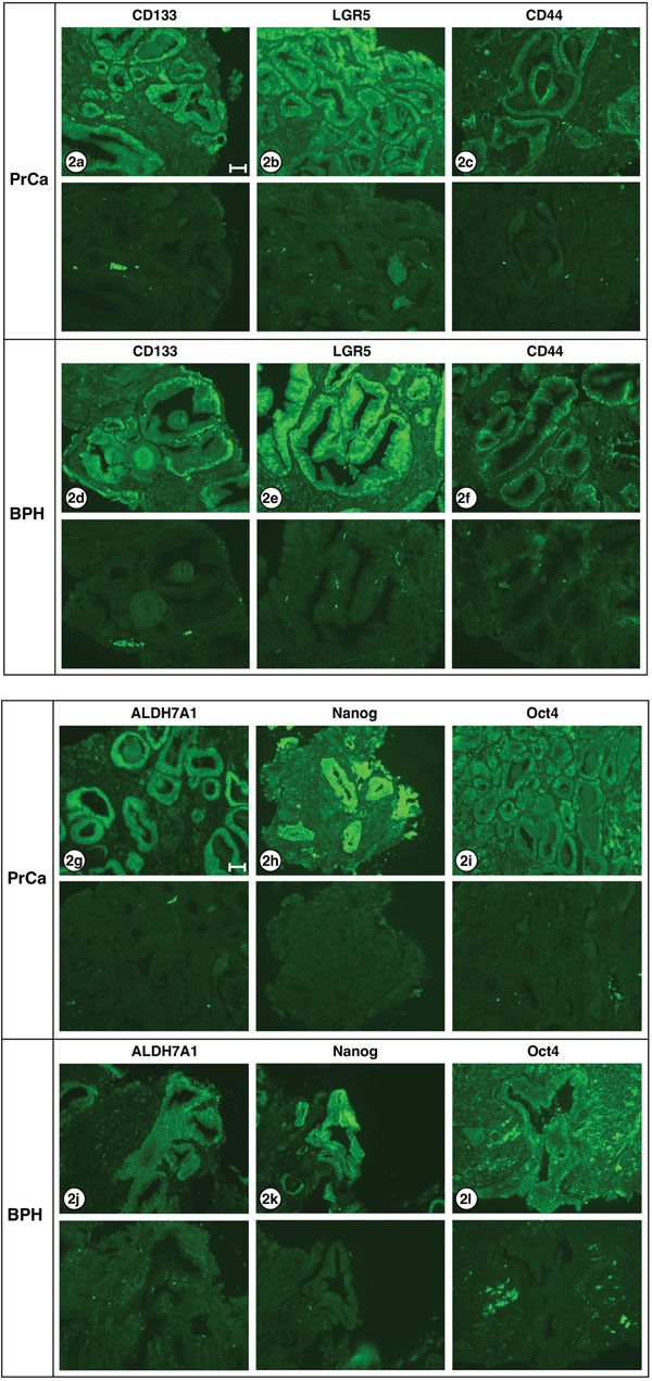
Figure 2. Stem cell markers expressed in PrCa and BPH.

Six different antibodies known to react with stem cells were examined for staining carcinoma and hyperplastic tissues: a, d. CD133; b, e. LGR5; c, f. CD44; g, j. ALDH7A1; h, k. Nanog; i, l. Oct4. The lower half of each frame shows the corresponding negative stain. The only differences in staining pattern were: CD133 appears to stain both cytoplasm and nucleus of carcinoma cells, but only the nucleus of hyperplastic cells. Oct4 stains both cytoplasm and nucleus of carcinoma cells, but only the cytoplasm of hyperplastic cells. ALDH7A1 shows weak nuclear staining and strong cytoplasmic staining in adenocarcinoma cells, but shows no detectable nuclear staining in hyperplastic cells. All exposures were 3 sec except ALDH7A1 and Nanog which were 1 sec. Images shown are from the following patient biopsie sections: 2a, A1; 2b, K1; 2c, C1; 2d, A1; 2e, K1; 2f, E1; 2g, C1; 2h, F1; 2i, C1; 2j, K1; 2k, C1; 2l; J1. Bar = 50 μm.
