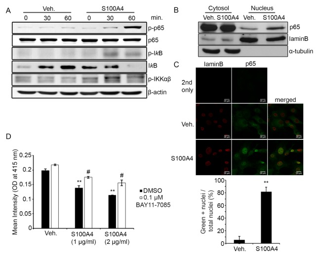
Fig. 3.
S100A4 induced NF-κB activation in osteoblasts. (A) Mouse calvarial preosteoblasts were serum-starved for five hours, stimulated with either a vehicle (Veh.) or recombinant mouse S100A4 (2 μg/ml) for the indicated time and subjected to Western blotting to detect protein levels of phosphorylated p65 (p-p65), total p65, phosphorylated IκB (p-IκB), total IκB, and phosphorylated IKKαβ. β-Actin is shown as a loading control. (B) Calvarial cells were serum-starved for five hours and stimulated with either vehicle or S100A4 (2 μg/ml) for one hour. Cytosolic proteins (30 μg) and nuclear proteins (8 μg) were separated and subjected to Western blotting to detect protein levels of p65, laminB, and α-tubulin. (C) Calvarial cells were serum-starved for five hours and stimulated with either a vehicle or S100A4 (2 μg/ml) for one hour. Cells were stained with anti-laminB (red) and anti-p65 (green). LaminB was labeled to locate the nuclear membrane. Cells were subjected to confocal microscopy and representative images displayed (top). Green positive (+) nuclei were counted and depicted as a graph (bottom). Primary antibodies were not added for 2nd-only samples. (D) Mouse calvarial preosteoblasts were cultured with a vehicle or S100A4 (1 or 2 μg/ml) in osteogenic medium for nine days, together with DMSO or indicated concentrations of BAY11-7085. Cells were subjected to Alizarin-red staining. The intensity of Alizarin-red stain was quantified after solubilization using cetylpyridium chloride. Error bars represent the SD of mean values. **P < .01 versus Veh.. #P < .01 versus DMSO.

