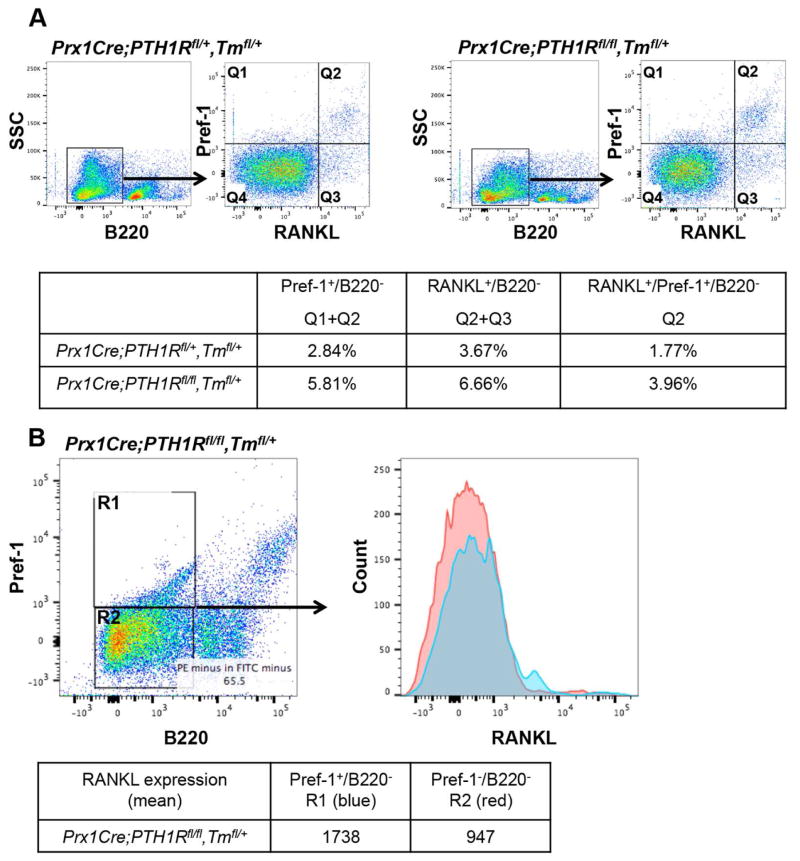
Figure 6. Loss of PTH1R in MSCs increases the number of RANKL expressing pre-adipocytes.
Bone marrow cells of 3-wk-old controls (Prx1Cre;PTH1Rfl/+,Tmfl/+, PTH1Rfl/fl) and mutants (Prx1Cre;PTH1Rfl/fl,Tmfl/+, Prx1Cre;PTH1Rfl/fl) were stained with FITC-anti-B220 antibody, PE-anti-Pref-1 and APC/Cy7-anti-RANKL antibody and analyzed by flow cytometry. (A) Cells were gated on B220− cells and characterized with Pref-1 and RANKL (separated into Q1–Q4). Q1+Q2 represent Pref1+B220− population, Q2+Q3 represent RANKL+B220− population and Q2 represents Pref-1+RANKL+B220− population. Table shows the percentage of each population. (B) Mutant cells were gated on B220 and Pref-1 and separated into R1 and R2, and each fraction was analyzed with RANKL expression. R1 represents Pref-1+RANKL+B220− and R2 represents Pref-1-RANKL+B220− population. Table shows mean fluorescence intensity of RANKL expression. Results shown are representative of three independent experiments displaying similar results.

