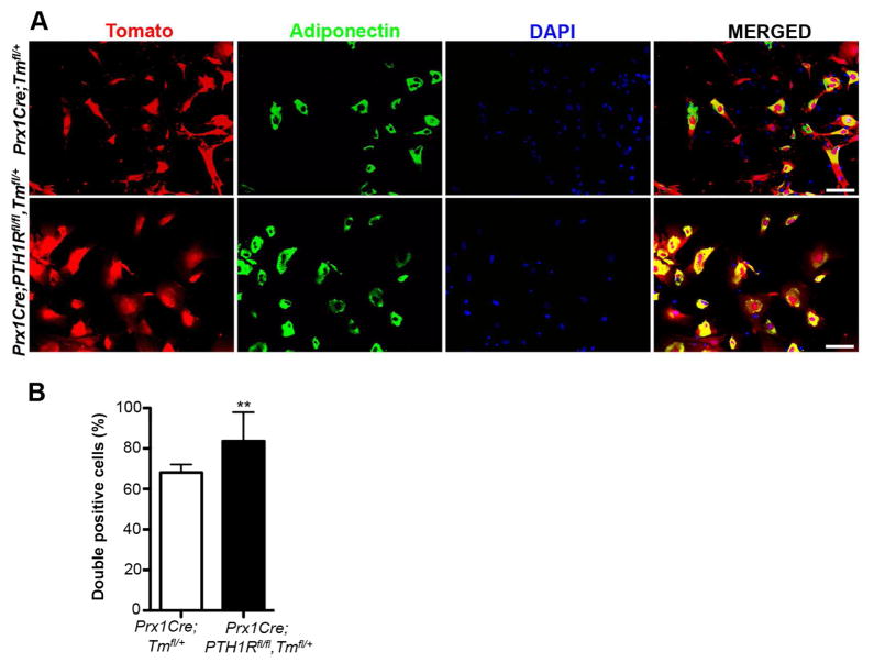
Figure 7. Cells ablated for PTH1R preferentially developed into adipocytes in vitro.
(A) Immunofluorescence staining of BMSCs cultured under adipogenic conditions derived from 3wk old Prx1Cre;Tmfl/+ and Prx1Cre;PTH1Rfl/fl,Tmfl/+ mice. Red: Prx1-Tomato positive cells, green: adipocytes expressing adiponectin, blue: DAPI stain for cell nuclei and yellow: double positive (red/green) cells rendering BMSCs undergoing adipogenic differentiation. Scale bar: 100μm. (B) The percentage of double positive cells in Prx1Cre;PTH1Rfl/fl,Tmfl/+ mice was significantly higher than in controls. n=3. **p<0.01 versus control. All graphs show mean ± SEM.

