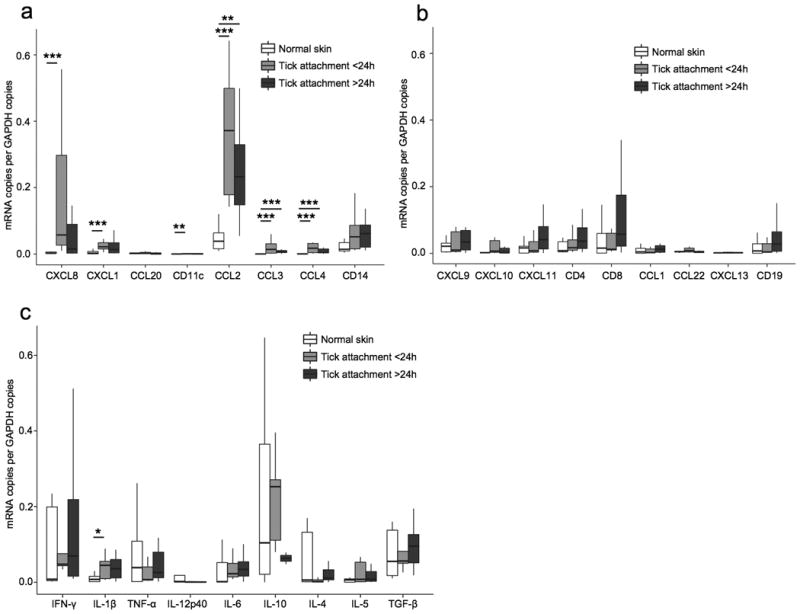
Figure 4. mRNA expression in skin lesions with a tick attachment period of <24 h (n=10) versus >24 h (n=8) compared to normal skin (n=9).

The same samples as in figure 3 are depicted. mRNA expression was measured by quantitative real-time PCR and expressed relative to the copies of glyceraldehyde-3-phosphate dehydrogenase (GAPDH). (a) Neutrophil chemoattractants (CXCL8, CXCL1), the dendritic cell chemoattractant CCL20, the dendritic cell marker CD11c, macrophage chemoattractants (CCL2, CCL3, CCL4) and the macrophage cell marker CD14. (b) Th1 cell chemoattractants (CXCL9, CXCL10, CXCL11), T cell markers (CD4, CD8), Th2 cell chemoattractants (CCL1, CCL22), the B cell chemoattractant CXCL13, and the B cell marker CD19. (c) Pro-inflammatory cytokines (IFN-γ, IL-1β, TNF-α, IL-12p40, IL-6) and anti-inflammatory cytokines (IL-10, IL-4, IL-5, and TGF-β). * P ≤ 0.05, ** P ≤ 0.01, *** P ≤ 0.001.
