
Figure 4. Comparative models of 8 Ig-like domains and comparison to an Ig-like domain in human Nup210.
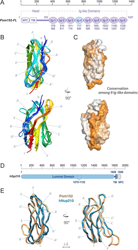
(A) Domain organization of Pom152FL and its domains drawn to scale. The head region contains the domain that faces the NPC inner ring (NPC) and the transmembrane segment (TM), followed by the lumenal domain composed of nine Ig-like repeats. The amino acid boundaries for each Ig-like repeat are indicated below them. The repeat analyzed by NMR (Ig-4, Pom152718–820) is highlighted in light blue.
(B) Comparative models of the remaining 8 Ig-like domains were built using the Pom152718–820 NMR structure as the template, followed by superposing them on the Pom152718–820 structure. The 8 domains share statistically significant sequence alignments to Pom152718–820 (E-values ranging from 3.3×10−49 to 6.8×10−39).
(C) Sequence conservation among the 9 Ig-like domains was mapped on the structure of Pom152718–820 using ConSurf (Ashkenazy et al., 2016). Low conservation, white; high conservation, orange.
(D) Domain organization of human Nup210. See legend of Figure 1A.
(E) Superposition of comparative models for yeast Pom152718–820 (orange) and human Nup2101079–1152 (blue).
See also Figure S3.