FIGURE 3.

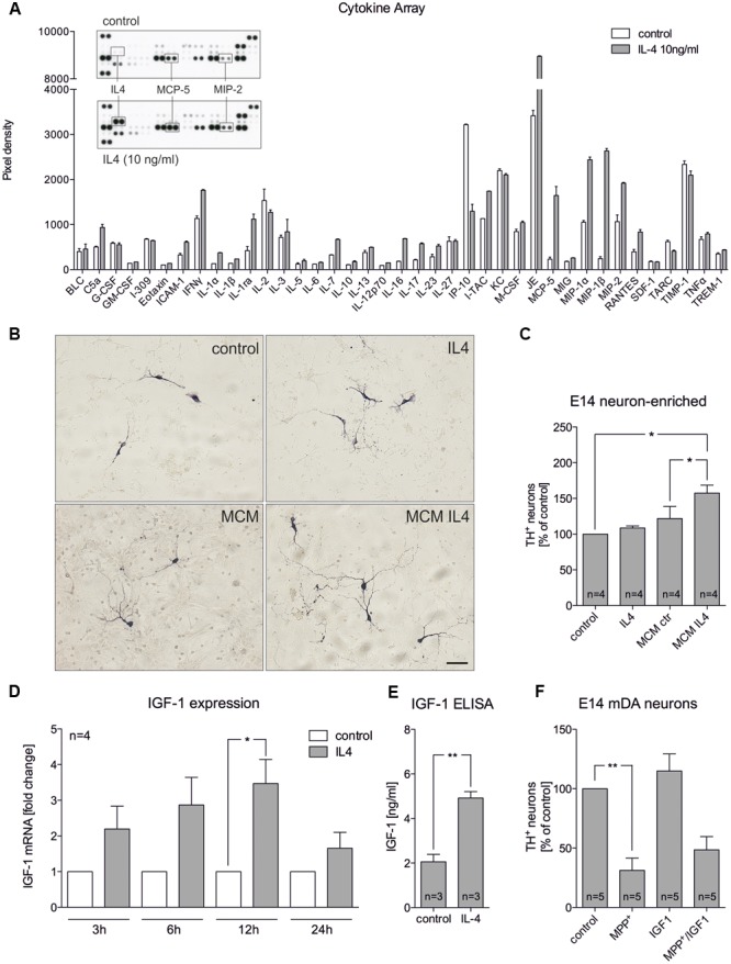
Microglia-conditioned medium after IL4 treatment is neuroprotective in vitro. (A) Treatment of primary microglia with recombinant IL4 (10 ng/ml) for 24 h results in modest changes of secreted chemokines and cytokines. As expected, IL4 was detected in IL4-treated samples and thus was not included in the densitometric spot analysis. Levels of MCP-5 and MIP-2 were increased after IL4 treatment. Data are given as mean ± SEM from two independent experiments. (B) E14 ventral midbrain neuron-enriched cultures were treated for 2 days with serum-free medium (control) and IL4 (10 ng/ml) or with microglia conditioned medium obtained after treatment of primary microglia for 24 h with serum-free medium (MCM) and IL4 at 10 ng/ml (MCM IL4). Scale bar indicates 50 μm. (C) Quantifications of TH+ neurons indicate that microglia conditioned medium after treatment with IL4 (MCM IL4) significantly increased neuron survival. IL4 alone was not able to promote neuroprotection. (D) IL4 treatment increases IGF-1 expression in primary microglia. After treatment for 3, 6, 12, and 24 h IGF-1 expression was determined using qPCR and is presented as fold change compared to untreated control cultures. Significant increases in IGF-1 expression was observed after 12 h. (E) IGF-1 secretion under control conditions and 24 h after treatment with IL4 (10 ng/ml) was detected using an IGF-1 ELISA. (F) Recombinant IGF-1 (50 ng/ml) increased neuron survival in E14 ventral midbrain neuron-enriched cultures without reaching significance. Data are given as mean ± SEM from four (B–D), three (C, ELISA) and five (E) independent experiments performed in duplicates. P-values derived from student’s t-test (D) are ∗p < 0.05 and ∗∗p < 0.01. P-values derived from one-way ANOVA followed by Bonferroni’s multiple comparison post-test are ∗p < 0.05 and ∗∗p < 0.01.
