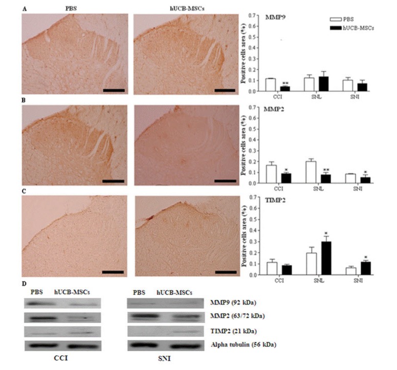
Fig. 3. Immunohistochemistry and western blot analysis for MMP-9, MMP-2, and TIMP-2 in the spinal cord.
The laminae I-II layers of the ipsilateral L4-L5 spinal dorsal horns and L4-L5 spinal cord in CCI and SNL models were dissected and sampled on 4 weeks, while those layers and that spinal cord in SNI models dissected on 8 weeks after transplantation of hUCB-MSCs. The cells positive for MMP-9, MMP-2, and TIMP-2 in the laminae I-II layers of the ipsilateral L4-L5 spinal dorsal horn were analyzed in the three neuropathic pain models. Positive cells area (%) presents the mean percentages of MMP-9 or MMP-2 or TIMP-2 immunoreactive (IR) neurons relative to the total number of neurons in the laminae I-II layers of the ipsilateral L4-L5 spinal dorsal horns. Expression of MMP-9 was only reduced in CCI model. In all of the neuropathic pain models, expression of MMP-2 was significantly decreased in the hUCB-MSCs group (n=11 in CCI, n=10 in SNL, n=12 in SNI) compared to control group (n=6 in CCI, n=7 in SNL, n=6 in SNI). In the hUCB-MSCs group, TIMP-2-positive cells were significantly increased in SNL and SNI models. Results for expression of those antibodies was similar in the western blot analysis. Statistical significance of difference was determined by Duncan's t-test. A p<0.05 was regarded as statistically significant. The data are presented as mean±S.E.M. *p<0.05, **p<0.01 indicate a significant difference compared to control group. The micrographs A and B are taken from the CCI group and C from the SNL group. A: MMP-9, B: MMP-2, C: TIMP-2, D: western blot analysis. Scale bars, 200 µm.

