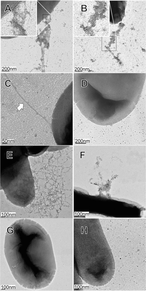
FIG 4.

Transmission electron microscopy of Bcf- and Stf-fimbriated bacteria. (A) E. coli AAEC189 pLDBAD-Stf, after Stf induction. The inset is a close-up image of fimbriae and fimbrial aggregates at the bacterial cell surface. (B) Immunogold-labeled Stf fimbriae and fimbrial aggregates on E. coli SE5000 containing pLDBAD-Stf using adsorbed rabbit anti-Stf antisera, as described in Materials and Methods, followed by anti-rabbit antibodies conjugated to 10-nm-diameter gold particles. The inset is a close-up image showing numerous gold particles attached to fimbrial aggregates. (C) E. coli AAEC189 pBAD33 (empty vector control). The white arrow denotes a flagellum (flagella were absent from SE5000). (D) Immunogold labeling of E. coli SE5000 pBAD33, as described for panel B. (E) E. coli AEEC189 pLDHSG-Bcf-S after Bcf induction. (F) Immunogold-labeled strain AJB4ΔbcfC pLDHSG-Bcf-L processed with adsorbed rabbit anti-Bcf antisera followed by anti-rabbit antibodies conjugated to 20-nm-diameter gold particles, showing entangled fimbriae with numerous gold particles attached. (G) E. coli SE5000 pHSG576 (empty vector control). (H) Immunogold labeling of strain AJB4ΔbcfC pHSG576, as described for panel F.
