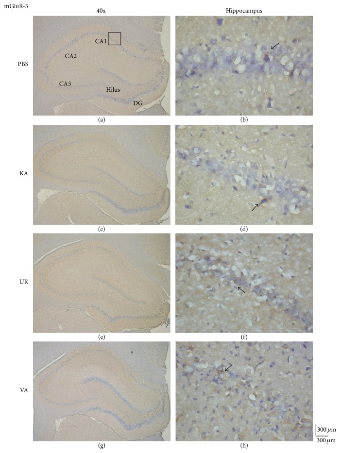
Figure 5.
Immunohistochemistry staining of mGluR3 and hematoxylin and eosin (HE) staining in hippocampal sections from the PBS, KA, UR, and VA-pretreated groups. HE (blue) and mGluR3 (brown) immunostaining in the whole hippocampus (a) and CA1 (b) in the PBS group. HE (blue) and mGluR3 (brown) immunostaining in the whole hippocampus (c) and CA1 (d) areas in the KA group. HE (blue) and mGluR3 (brown) immunostaining in the whole hippocampus (e) and CA1 (f) areas in the UR group. HE (blue) and mGluR3 (brown) immunostaining in the whole hippocampus (g) and CA1 (h) areas in the VA group. The left panel was imaged at 40x magnification, and the right panel was imaged at 400x magnification.

