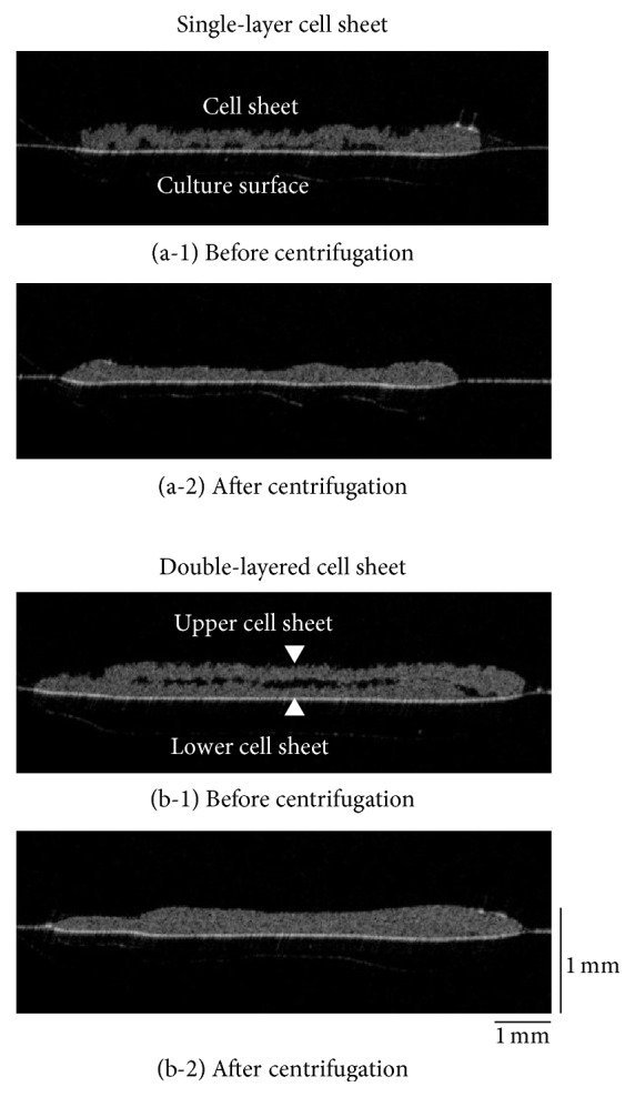
Figure 2.

Observation of double-layered human iPS cell-derived cardiac cell sheets by optical coherence tomography (OCT). Just after the transfer of a human iPS cell-derived cardiac cell sheet onto a polystyrene culture dish, there were numerous spaces between the surfaces (a-1). After centrifugation, the spaces were almost entirely eliminated (a-2). Just after the transfer of a second cell sheet onto the first cell sheet, again there were numerous spaces between the surfaces (b-1). After centrifugation, the spaces were almost entirely eliminated (b-2). Three experiments were performed independently and all of them showed similar results. An OCT system [Proto 3(DT)] was used in this study.
