Fig. 10.

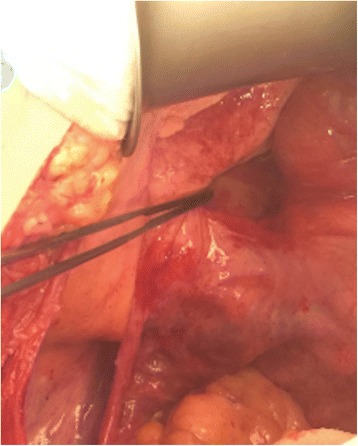
Intraoperative view of pericecal and retrocecal mucin. The disease was limited to the right iliac fossa. Pathological analysis analysis revealed no cells in the mucin

Intraoperative view of pericecal and retrocecal mucin. The disease was limited to the right iliac fossa. Pathological analysis analysis revealed no cells in the mucin