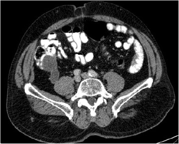
Fig. 8.

Axial computerized tomography view of a burst cecal appendix (up to 3.5 cm), with thin and regular walls, and no signs of densification of adjacent adipose tissue. This corresponds to the cystic formation already described in the ultrasound, compatible with mucocele of undetermined etiology. Appendectomy revealed well-differentiated mucinous adenocarcinoma, with invasion into of the muscularis mucosae
