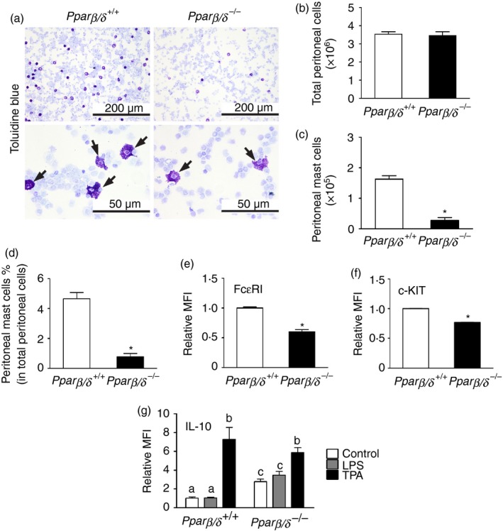
Figure 4.

Peroxisome proliferator‐activated receptor‐β/δ (PPAR β/δ) is required for maturation of peritoneal mast cells (PMCs). (a) Representative photomicrographs of cytospun peritoneal cells from adult Pparβ/δ +/+ and Pparβ/δ −/− mice stained by toluidine blue. Arrowheads indicated toluidine blue‐positive mast cells. Upper panels: Magnification × 20. Bar = 200 μm. Lower panels: Magnification × 40. Bar = 50 μm. The numbers of (b) total peritoneal cells and (c) PMCs were counted, and (d) the average percentage of mast cells in total peritoneal cells was determined. Relative mean fluorescent intensities (MFI) of (e) Fcε RI and (f) c‐KIT of PMCs were determined by flow cytometry. Values represent the mean ± SEM. *Significantly different from Pparβ/δ +/+, P ≤ 0·05. (g) Peritoneal cells from adult Pparβ/δ +/+ and Pparβ/δ −/− mice were treated with lipopolysaccharide (LPS) or 12‐O‐tetradecanoylphorbol‐13‐acetate (TPA) for 24 hr, and expression of interleukin‐10 was determined by flow cytometry. Values represent the mean ± SEM. Values with different superscript letters are significantly different at P ≤ 0·05. [Colour figure can be viewed at wileyonlinelibrary. com]
